 Autonomic dysreflexia can be reversed using epidural electrical neuromodulation
A 53-year-old man living with a chronic spinal cord injury wakes in the middle of the night with a crushing headache. His sheets are soaked. He is trembling with chills, and something feels profoundly wrong. His caregiver takes his blood pressure; it reads 190/120 mm Hg. “I think you’re having a stroke,” his caregiver tells him. But this is not a stroke. It is autonomic dysreflexia, a sudden hypertensive crisis and one of the most dangerous yet underrecognized complications of spinal cord injury.
To the public, spinal cord injury is synonymous with paralysis because it is the most visible manifestation of spinal cord injury. With the loss of brain control, this condition also causes broad disruption of autonomic regulation over all the physiological functions that keep the body in balance, including breathing, blood pressure, bowel control, bladder function, and sexual function. This disconnection of the spinal cord from the rest of the nervous system also causes widespread and uncontrollable anatomical reorganization. Spinal cord neurons that were once controlled by brainstem centers become hyperactive, and routine sensory inputs that normally pass unnoticed can trigger massive neuronal surges with far-reaching physiological consequences. Autonomic dysreflexia is the clinical manifestation of this hyperactivity. It might begin with a headache or goosebumps, but it can escalate to a stroke or even death.
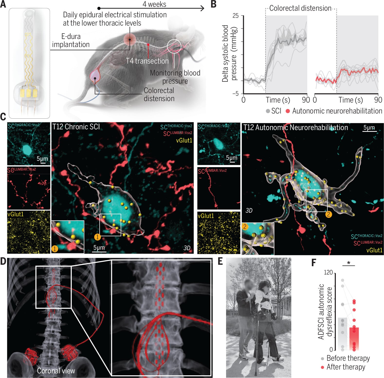
Autonomic neurorehabilitation abolished autonomic dysrefl exia in mice and reduced its severity in humans with chronic spinal cord injury.
(A) Schematic of autonomic neurorehabilitation and the paradigm used to quantify autonomic dysrefl exia severity.
(B) Pressor responses (left; individual mice and mean trace) and autonomic dysrefl exia severity (right) in mice with chronic spinal cord injury (SCI) and mice that underwent autonomic neurorehabilitation for 4 weeks starting 1 week after SCI.
(C) Immunofluorescence images show vGLUT1+synaptic puncta and synaptic-like appositions from lumbosacral Vsx2 (SCLUMBAR::Vsx2) neurons onto lower thoracic Vsx2 (SCTHORACIC::Vsx2) neurons in mice with SCI and mice that underwent autonomic neurorehabilitation.
(D) Sagittal and coronal reconstructions from postoperative computed tomography (CT) scans show a participant’s spinal cord implant.
(E) Use of epidural electrical stimulation programs to improve blood pressure instability during upright rehabilitation.
(F) Autonomic dysfunction following spinal cord injury (ADFSCI) autonomic dysrefl exia scores before implantation and 6 months to 2 years after implantation and daily use of the system to regulate blood pressure. *P = 0.02118.
During my graduate studies, I worked with people with high-level spinal cord injury who experienced severe autonomic dysreflexia. I saw firsthand how frightening and debilitating these episodes can be. I observed that clinicians and patients can sometimes stop a crisis after it starts by removing the triggering stimulus, yet they have no way to prevent the next one from occurring. This is because, until recently, the hyperactive neurons responsible for these crises remained unidentified and therefore could not be targeted by any therapy.
My determination to understand this dangerous clinical condition led me and my colleagues to uncover the neuronal architecture underlying autonomic dysreflexia. We then developed a neuromodulatory therapy to target this architecture that prevents hypertensive episodes before they begin by using neuroscience approaches we developed to investigate blood pressure regulation.
We began by mapping which parts of the spinal cord become hyperactive during an episode of autonomic dysreflexia. In chronically injured mice, I quantified activity throughout the spinal cord using cFos labeling and found the strongest activation in the lower thoracic and lumbosacral segments. To reveal which cell types were driving the response, I turned to single-cell transcriptomics to pinpoint the neuronal subtypes associated with autonomic dysreflexia. I compared spinal cords from chronically injured mice in which autonomic dysreflexia was induced with those of injured controls only. Unexpectedly, analyses of both the lower thoracic and lumbosacral regions converged on an excitatory neuronal subpopulation expressing the transcription factor Vsx29, which are referred to as Vsx2 neurons. These neurons have been linked to motor functions, but their involvement in blood pressure control had not yet been described. To explain this, I hypothesized that their ability to modulate blood pressure emerges only after spinal cord injury. Consistent with this idea, removal of Vsx2 neurons from either region prevented autonomic dysreflexia in injured mice yet had no measurable effect on blood pressure in uninjured animals.
Next, I aimed to understand how these Vsx2-expressing neurons cause autonomic dysreflexia after spinal cord injury. To address this question, I mapped the axonal projection patterns of Vsx2 neurons in the lumbosacral and lower thoracic spinal cord. Strikingly, I found that after injury, Vsx2-expressing neurons underwent a massive anatomical reorganization, whereby these neurons located in the lumbosacral spinal cord propelled dense projections to the Vsx2 neurons in the lower thoracic spinal cord. These neuronal projections, however, were absent in uninjured mice.
Once I understood how this neuronal architecture changes, I asked whether neuromodulation could be a therapeutic solution to this long-standing problem. Vsx2 neurons have been therapeutically modulated using neurostimulation to alter neuronal activity patterns, particularly in the recovery of walking. I wondered whether Vsx2 neurons in the thoracic spinal cord could be a therapeutic entry point for autonomic dysreflexia. I found that in mice, a 1-month regimen of autonomic neurorehabilitation involving daily spinal cord stimulation sessions abolished autonomic dysreflexia in every animal tested (see the figure, panels A and B). Moreover, I elucidated the mechanism underlying this improvement. Because Vsx2 interneurons link the projections that cause autonomic dysreflexia to the pathway that epidural electrical stimulation accesses to control blood pressure, repeated stimulation retrains the spinal cord. Over time, this stimulation shifted reorganization toward steadier sympathetic output and shut down the maladaptive anatomical reorganization that triggers autonomic dysreflexia (see the figure, panel C).
These remarkable findings in mice compelled me to ask whether autonomic dysreflexia could be targeted with similar approaches in humans with spinal cord injury. I leveraged data from ongoing clinical trials to quantify autonomic dysreflexia symptoms in participants who used epidural stimulation to manage their blood pressure. These participants reported significantly fewer headaches and heart palpitations, two of the most prominent symptoms of autonomic dysreflexia (see the figure, panels D to F). My clinical observations triggered the inclusion of autonomic dysreflexia as a key outcome measure in a new multisite pivotal clinical trial, Empower BP run by ONWARD Medical, that will assess the safety and efficacy of epidural electrical spinal cord stimulation for the modulation of blood pressure in people with spinal cord injury.
I imagine a world in which that same 53-year-old man wakes in the night and pauses, not because he is terrified, but because he recognizes an early warning sign. He may still feel a hint of pressure behind his eyes, but the crushing headache does not arrive. His blood pressure no longer surges into danger. The sensations that once mimicked a stroke have become something else entirely. They tell him his bladder is full or that something below the level of injury needs attention. He knows that the consistent stimulation from his spinal cord implant has reshaped his nervous system to prevent dangerous elevations of blood pressure. Autonomic dysreflexia is no longer an unpredictable crisis that controls him, but a manageable message from a spinal cord whose neuronal architectures have been rewired.
2026 GRAND PRIZE WINNER of the Science & PINS Prize
 Jan Elaine Soriano
Jan Elaine Soriano received an undergraduate degree from Mount Royal University and a PhD from the University of Calgary. She is presently a postdoctoral fellow at École Polytechnique Fédérale de Lausanne. Her research focuses on leveraging single-cell sequencing, genetic manipulation, and neuromodulation to develop therapeutics for people with neurological conditions. www.science.org/doi/10.1126/science.aej5900